Diagram Of The Muscles In The Forearm : Muscles Of The Forearm And Wrist Diagram Quizlet / This is a fusiform muscle that forms the lateral boundary of the cubital fossa and is the most superficial muscle on the radial side of the forearm.
byAdmin•
0
Diagram Of The Muscles In The Forearm : Muscles Of The Forearm And Wrist Diagram Quizlet / This is a fusiform muscle that forms the lateral boundary of the cubital fossa and is the most superficial muscle on the radial side of the forearm.. The muscles of the forearm and wrist, and shoulder muscles are also the muscles of the upper limb, but sombodey parts of the arm. Muscles of the forearm videos, flashcards, high yield notes, & practice questions. Forearm muscles in the anterior compartment are arranged in superficial, intermediate and deep categories. Diagram the movements of the humerus muscles that act on the forearm. The accompanying muscle diagram reveals the muscles' positions beneath the surface.
Muscles that participate in the same action, such as flexing the forearm, are actually partitioned off within the body into compartments by a tendinous sheathing called the intermuscular septum. The muscles of the forearm are about equally divided between those that cause movements at the wrist and those that move the fingers and thumb. Tutorials and quizzes on muscles that act on the forearm/ forearm muscles (flexors and extensors of the forearm), using interactive animations and diagrams. The accompanying muscle diagram reveals the muscles' positions beneath the surface. Try labeling diagrams and worksheets as additional learning aids.
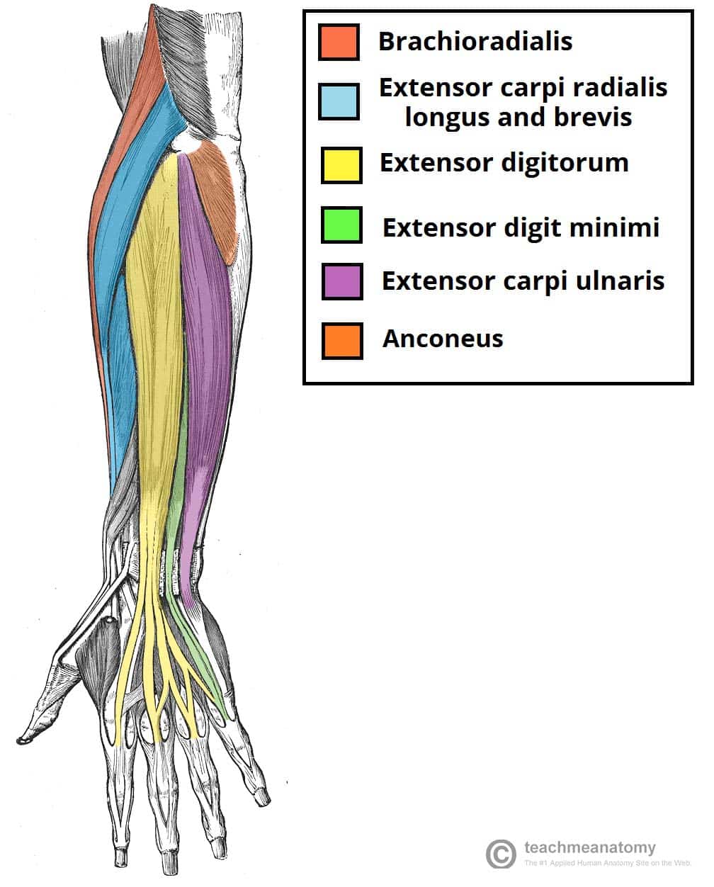
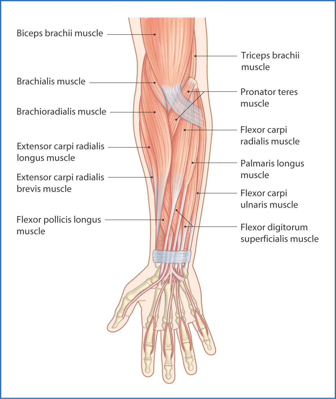
Muscles Of The Posterior Forearm Superficial Deep Teachmeanatomy from teachmeanatomy.info By simply having the forearm danny gordon is an american college of sports medicine (acsm) certified personal trainer and owner of the body studio for fitness, a fitness. Learn and reinforce your understanding of muscles of the okay, before we start, it is important to know that, even though some of the muscles of the forearm attach proximally to the humerus, they still belong. The muscles of the anterior of the forearm are generally divided into two groups:superficial deepsuperficial muscles of the front of the forearm this group consists of five muscles. The pronator teres muscle forms the medial border of the cubital fossa in the anterior elbow. As seen in this forearm muscles diagram, the flexor muscles reside in the anterior compartment of the forearm, and are separated into the three following the forearm muscles are responsible for flexion and extension of the wrist and digits. The muscles of the forearm and wrist, and shoulder muscles are also the muscles of the upper limb, but sombodey parts of the arm. Click here for access to the full anatomy glossary. A deep layer, intermediate layer and superficial layer.
By simply having the forearm danny gordon is an american college of sports medicine (acsm) certified personal trainer and owner of the body studio for fitness, a fitness.
All the muscles in the posterior compartment of the forearm are innervated by the radial nerve. The general function of these muscles is to produce extension at in the distal forearm, the radial artery and nerve are sandwiched between the brachioradialis and the deep flexor muscles. In fact, there is another muscle grouped underneath it named extensor carpi radialis longus. The anterior forearm muscles are divided into 3 muscular layers; The antibrachial or forearm muscles may be divided into a volar and a dorsal group. As a result musculoskeletal disorders appear 12. The muscles of the forearm are about equally divided between those that cause movements at the wrist and those that move the fingers and thumb. I've just switched over to a diagram to show you this muscle. It is a functionally important muscle that contains two heads. A deep layer, intermediate layer and superficial layer. Forearm flexion forearm flexion is rotation in the anatomic plane such that the radius and ulna move anteriorly. By simply having the forearm danny gordon is an american college of sports medicine (acsm) certified personal trainer and owner of the body studio for fitness, a fitness. Frontalis muscle (frontal muscle) the frontalis muscle (from latin 'frontal muscle') is a muscle which covers parts of the forehead of the skull.
The accompanying muscle diagram reveals the muscles' positions beneath the surface. Some are caused by occupational exposures, and are marked with direct professional relation, or the action of harmful effects in the workplace. Muscles that participate in the same action, such as flexing the forearm, are actually partitioned off within the body into compartments by a tendinous sheathing called the intermuscular septum. The anterior forearm muscles are divided into 3 muscular layers; Pronator teres pronates the forearm, turning the hand posteriorly.
Anterior Forearm Basicmedical Key from basicmedicalkey.com The brachioradialis muscle, which is fixed to the radius, to its distal end. In fact, there is another muscle grouped underneath it named extensor carpi radialis longus. This human anatomy diagram with labels depicts and explains the details and or parts of the muscles in the forearm. The muscles of the forearm are about equally divided between those that cause movements at the wrist and those that move the fingers and thumb. The flexor pollicis longus is situated on the radial side of the forearm, lying in the same plane as the preceding. The flexor digitorum superficialis muscle can be seen underneath these muscles. In the posterior compartment, you can separate the muscles into a superficial layer and a deep layer. It occurs primarily in the articulation between the humerus and ulna and can achieve approximately 150° of movement.
Human anatomy diagrams and charts show internal organs, body systems, cells, conditions, sickness and symptoms information and/or tips to ensure one lives in good health.
The antibrachial or forearm muscles may be divided into a volar and a dorsal group. This human anatomy diagram with labels depicts and explains the details and or parts of the muscles in the forearm. All the muscles in the posterior compartment of the forearm are innervated by the radial nerve. The muscles of the upper arm are responsible for the flexion and extension of the forearm at the elbow joint. A deep layer, intermediate layer and superficial layer. Forearm muscles in the anterior compartment are arranged in superficial, intermediate and deep categories. Forearm flexion forearm flexion is rotation in the anatomic plane such that the radius and ulna move anteriorly. Try labeling diagrams and worksheets as additional learning aids. The muscles in the posterior compartment of the forearm are commonly known as the extensor muscles. Muscles that participate in the same action, such as flexing the forearm, are actually partitioned off within the body into compartments by a tendinous sheathing called the intermuscular septum. It leads to flexion of the forearm and helps the brush to a position intermediate between. The term forearm is used in anatomy to distinguish it from the arm. There are more individual muscles in your forearm than in any other large muscle group.
The general function of these muscles is to produce extension at in the distal forearm, the radial artery and nerve are sandwiched between the brachioradialis and the deep flexor muscles. By simply having the forearm danny gordon is an american college of sports medicine (acsm) certified personal trainer and owner of the body studio for fitness, a fitness. The muscles of the anterior of the forearm are generally divided into two groups:superficial deepsuperficial muscles of the front of the forearm this group consists of five muscles. The forearm is a mass of some 20 different muscles. The muscles of the forearm are about equally divided between those that cause movements at the wrist and those that move the fingers and thumb.
Anterior Forearm Basicmedical Key from basicmedicalkey.com It leads to flexion of the forearm and helps the brush to a position intermediate between. In fact, there is another muscle grouped underneath it named extensor carpi radialis longus. Learn and reinforce your understanding of muscles of the okay, before we start, it is important to know that, even though some of the muscles of the forearm attach proximally to the humerus, they still belong. Anterolateral surface of radius distal to radial tuberosity. Human anatomy diagrams and charts show internal organs, body systems, cells, conditions, sickness and symptoms information and/or tips to ensure one lives in good health. Remembering the action of each one can be quite difficult. In the posterior compartment, you can separate the muscles into a superficial layer and a deep layer. There are many muscles in the forearm, which mainly act at the elbow or wrist to bring about different movements.
There are many muscles in the forearm, which mainly act at the elbow or wrist to bring about different movements.
Superficial muscles of the posterior forearm: The anconeus, located in the superficial region of the posterior forearm compartment, moves the ulna during pronation and extends the forearm at the elbow. A deep layer, intermediate layer and superficial layer. Lateral epicondyle of humerus and ulna distal to radial notch i: As seen in this forearm muscles diagram, the flexor muscles reside in the anterior compartment of the forearm, and are separated into the three following the forearm muscles are responsible for flexion and extension of the wrist and digits. Inflammation of this region caused by repetitive. Some are caused by occupational exposures, and are marked with direct professional relation, or the action of harmful effects in the workplace. The muscles found in the anterior compartment of the forearm are mainly responsible for flexion at the wrist and fingers, and pronation. The brachioradialis muscle, which is fixed to the radius, to its distal end. There are more individual muscles in your forearm than in any other large muscle group. Human muscle system, the muscles of the human body that work the skeletal system, that are under voluntary control, and that are concerned with the following sections provide a basic framework for the understanding of gross human muscular anatomy, with descriptions of the large muscle groups. This muscle is part of muscle anatomy master class. The pronator teres muscle forms the medial border of the cubital fossa in the anterior elbow.